Read protocols for the treatment of gynecological patients in the clinic. Is IVF done for uterine fibroids?
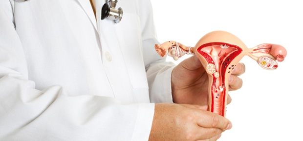
uterine fibroids is a benign hormone-dependent tumor that develops from the myometrium (the muscular layer of the uterus). If earlier this tumor was detected in women over 40 years old, now it is found in 20% of women aged 30 years, and in some cases in 25-year-old patients.
Rarely enough, one fibroid node is found in the uterus, much more often there are several. Mostly fibroids are located in the body of the uterus, but sometimes they are located in the cervix. Myoma nodes can be located between the muscles of the uterus, and then they are called intermuscular, or interstitial. They can also be located on the surface of the uterus, and then they talk about subserous, or subperitoneal, nodes, and if the node protrudes into the uterine cavity, then it is called submucosal, or submucosal.
Causes of uterine fibroids
With uterine fibroids, myometrial cells begin to spontaneously divide. To date, it has not been established exactly why this happens, but it is known that this process stimulated by hormones. Excess estrogen in the body leads to the growth of fibroids, and progesterone, on the contrary, suppresses this process. But not always in women with uterine fibroids in the blood is determined by a high level of estrogens. This is due to the fact that local changes in the level of these hormones are not reflected or slightly changed in the blood.
Risk factors
- Late onset of menstruation (after 14-16 years).
- Frequent abortions.
- Gynecological diseases: chronic inflammatory diseases of the genital organs, endometriosis, etc.
- Violation of the system hypothalamus - pituitary - ovaries. Normally, the hypothalamus produces hormones that stimulate the production of follicle-stimulating (FSH) and luteinizing (LH) hormones in the pituitary gland. They, in turn, act on the ovaries, regulating their work and their production of the hormones estrogen and progesterone. Failure at one of the levels leads to hormonal disorders.
- hereditary predisposition.
Symptoms of uterine fibroids
Symptoms of uterine fibroids depend on the location of the nodes, their size, and the rate of growth of the tumor. In 50-60% of women, the disease can occur without any symptoms.
As the fibroid grows, the following manifestations of the disease occur:
- Violations menstrual cycle- the main and earliest symptom of uterine fibroids. Menstruation lasts 7 or more days, is quite plentiful, and sometimes after the end of menstruation, spotting may continue for some time. Bleeding can also occur between periods.
- Stomach ache. The pain is mainly in the lower abdomen, the lumbar region. Prolonged pain may indicate the rapid growth of the tumor. They are connected with the fact that growing nodes compress the nerves that pass in the small pelvis. Pain can be aching, pulling and occur throughout the entire menstrual cycle. There may be cramping pain during menstruation. They occur if the node protrudes into the uterine cavity.
- Violation of the function of adjacent organs - occurs with large sizes of uterine fibroids, when it compresses the bladder, rectum. Constipation and urinary problems may occur.
Diagnosis of uterine fibroids
An obstetrician-gynecologist can establish the diagnosis of uterine fibroids. Already conducting a vaginal examination, with a large tumor, the doctor determines an enlarged uterus with a bumpy surface.
For the diagnosis of uterine fibroids, instrumental research methods are used:
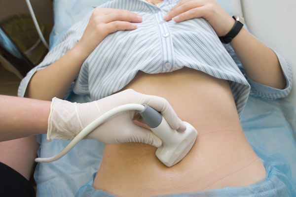
- Ultrasound of the uterus (the main diagnostic method);
- hysteroscopy (introduction into the uterus of a special device with a camera that allows you to examine the uterine cavity) - is performed if you suspect that the node protrudes into the uterine cavity (submucosal, or submucosal node);
- laparoscopy (introduction of a special camera into the abdominal cavity and examination of the uterus);
- magnetic resonance imaging (MRI) of the pelvic organs (in rare cases).
To characterize the size of uterine fibroids, gynecologists draw an analogy with pregnancy. For example, you can hear that fibroids correspond to 10 weeks of pregnancy. This means that the uterus due to fibroids is enlarged in the same way as at 10 weeks of pregnancy.
Diseases with similar symptoms
- Cystadenoma (cystoma) of the ovary
- Uterine sarcoma
- Tumor of the retroperitoneum
- Intestinal tumor
- ovarian cancer
Complications of uterine fibroids
- Anemia. It is due to the fact that during bleeding a woman loses a lot of blood, and with it iron, which is the main carrier of oxygen to all organs and tissues. In this case, the patient's condition worsens. She feels weakness, lethargy, drowsiness, dizziness, fainting, etc.
- Necrosis of the myomatous node. There is a violation of blood flow to the node, which provokes the death of its tissues. This can occur when the pedicle of the node located on the surface of the uterus is twisted. In this case, immediate surgery is required. If it is not carried out on time, peritonitis may develop.
- Degeneration of the myomatous node into a malignant tumor (up to 2% of cases).
- Reducing the likelihood of pregnancy, and during the expectation of a child, complications occur more often, which can also happen during childbirth.
Treatment of uterine fibroids
The choice of treatment for uterine fibroids depends on its size, the age of the woman and the desire to have children. There are two methods of treatment: conservative (the use of various drugs that slow down the growth of the tumor) and surgical.
Used for conservative treatment hormonal preparations. These are progesterone-based drugs, combined oral contraceptives, intrauterine device containing hormones, gonadoliberin agonists. The use of GnRH agonists reduces the size of fibroids by 55%. But after stopping treatment, uterine fibroids may start growing again.
Surgical treatment is used for large sizes of uterine fibroids, with heavy bleeding, severe pain, rapid tumor growth, malignant degeneration, etc.
In young women, only the fibroids are removed, and the uterus is preserved (myomectomy). The operation can be performed laparoscopically. At the same time, small incisions are made on the abdomen, an optical device is inserted into the abdominal cavity - a laparoscope and special instruments, with the help of which the nodes are removed.
If the uterine fibroid is large and it is impossible to remove it, then the uterus is removed with the preservation of the cervix or the entire uterus is removed.
Currently, modern methods of treatment of uterine fibroids are used:
- Embolization of the uterine arteries - a special substance is injected into the artery that supplies blood to the fibroid node, which clogs its lumen. After the blood stops flowing to the tumor, its necrosis (necrosis) occurs.
- Hysteroresectoscopy - a special optical device and instruments are inserted into the uterine cavity, and the fibroid node is removed with the help of current. The operation is possible if the node protrudes into the uterine cavity (submucosal fibroids).
Nota Bene!
If a woman with uterine fibroids becomes pregnant, then at first the tumor may grow, but this is not a reason for terminating the pregnancy. By 16-17 weeks, tumor growth slows down and even stops. During the period of expectation of a child with uterine fibroids, there is a risk of threatened miscarriage.After the onset of menopause, uterine fibroids do not occur, since the level of estrogen, the hormone that provokes the growth of this tumor, significantly decreases in the woman's body.
Prevention of uterine fibroids
- regular visits to the gynecologist once every six months;
- refusal of abortions and the use of hormonal contraceptives to prevent pregnancy.
Expert: Isaeva I. A., obstetrician-gynecologist
Prepared from:
- Gynecology: a national guide. Ed. V. I. Kulakov, G. M. Savelyeva, I. B. Manukhin. - M.: GEOTAR-Media, 2009.
- Women's consultation. Ed. V. E. Radzinsky. - M.: GEOTAR-Media, 2010.
- Strizhakov A. N. Minimally invasive surgery in gynecology. - M.: Medicine, 2001.
- Savitsky G. A. Uterine fibroids. - St. Petersburg: Way, 2000.
Uterine fibroids (MM) is a monoclonal hormone-sensitive proliferate consisting of phenotypically altered smooth muscle cells of the myometrium.
SYNONYMS OF UTERINE MYOMA
Leiomyoma, fibroma, fibromyoma.
ICD-10 CODE
D25 Leiomyoma of the uterus.
D25.0 Submucosal uterine leiomyoma.
D25.1 Intramural uterine leiomyoma.
D25.2 Subserous leiomyoma of the uterus.
D25.9 Uterine leiomyoma, unspecified.
EPIDEMIOLOGY OF uterine fibroids
MM is detected on average in 80% of women (according to autopsy studies). Clinically, MM manifests itself in 30–35% of women over the age of 35; twice as common in blacks.
PREVENTION OF uterine fibroids
A proven reduction in the risk of developing MM was noted with long-term use of combined oral contraceptives, in women who gave birth often, in women who did not undergo abortions and curettage of the uterine mucosa, who did not suffer from inflammatory diseases of the pelvic organs.
SCREENING
Ultrasound of the pelvic organs is performed once a year, starting from the age of 25.
CLASSIFICATION OF UTERINE MYOMAS
According to localization, the following types of fibroids are distinguished:
Topographic classification:
- submucosal nodes
♦ 0 type - myomatous node completely in the uterine cavity;
♦ Type I - less than 50% of the volume of the myomatous node is located intermuscularly, most of it is located in the uterine cavity;
♦ II type - more than 50% of the volume of the myomatous node is located intermuscularly, a smaller part of it is in the uterine cavity; - subserous nodes
♦0 type - myoma node on the leg, located completely in the abdominal cavity;
♦ Type I - less than 50% of the volume of the myomatous node is located intermuscularly, most of it is located in the abdominal cavity;
♦ II type - more than 50% of the volume of the myomatous node is located intermuscularly, a smaller part of it is located in the abdominal cavity.
Histological classification:
- simple;
- cellular;
- mitotically active;
- whimsical;
- atypical;
- lipoleiomyoma;
- epithelioid;
- hemorrhagic;
- vascular;
- myxoid;
- myoma with hematopoietic elements.
Clinical classification:
- clinically insignificant fibroids or small fibroids;
- small multiple uterine fibroids;
- uterine fibroids of medium size;
- multiple uterine fibroids with an average size of the dominant node;
- large uterine fibroids;
- submucosal uterine fibroids;
- uterine fibroids on the leg;
- complex uterine fibroids.
ETIOLOGY (CAUSES) OF uterine fibroids
There are two theories on the origin of the MM progenitor cell. One implies the appearance of a cell defect during the ontogenetic development of the uterus due to a long unstable period of embryonic smooth muscle cells, the second suggests the possibility of cell damage in a mature uterus. The fact that, according to autopsy studies, the prevalence of MM reaches 80%, allows us to consider the second theory of the origin of the progenitor cell more plausible.
PATHOGENESIS OF uterine fibroids
The formation of the growth germ of the myomatous node occurs as follows. It can be assumed that during the repeated cycles of myometrial hyperplasia during the menstrual cycle, smooth muscle cells accumulate, in which the apoptosis process is disrupted, and these proliferating cells are exposed to various damaging factors. Damage factors can be: ischemia due to spasm of the spiral arteries during menstruation, inflammatory process, traumatic impact during medical manipulations or a focus of endometriosis.
With each menstrual cycle, the number of damaged cells accumulates. Some of the cells are sooner or later eliminated from the myometrium, while others begin to form the rudiments of myomatous nodes with different potential for growth. The active growth germ in the first stages develops due to physiological fluctuations in the concentration of hormones during the menstrual cycle. Subsequently, the resulting cooperation of cells activates autocrine-paracrine mechanisms caused by growth factors, forms local autonomous mechanisms for maintaining growth (local production of estrogens from androgens and the formation of connective tissue), and the significance of physiological concentrations of sex hormones for the formation of a myomatous node ceases to be the main one.
The proliferative activity of MM cells is due to dysregulation of the HMGIC and HMGIY genes located on chromosomes 12 and 6, respectively, that is, in the loci of the most common chromosomal aberrations characteristic of this formation. The expression product of the HMGIY and HMGIC genes is recognized as proteins belonging to different families of the group of highly mobile proteins. Aberrant expression of HMGIC and HMGIY proteins most often characterizes the malignant process. At the same time, dysregulation of these proteins due to chromosomal rearrangements is most often detected in various benign mesenchymal formations. The pattern of expression of HMGIC and HMGIY proteins indicates their participation in the rapid growth of embryonic tissues and tissues in culture.
The monoclonal proliferation of smooth muscle cells of the myometrium, in which the program of clonal tissue proliferation is activated due to the dysregulation of the HMG genes, increases in size against the background of a normal hormonal background, while the cells of the unchanged myometrium are in a state of relative rest.
The value of the hormonal background for the growth of the myomatous node up to a certain stage is critical. With an increase in size, the formation of autocrine-paracrine regulation of growth and the formation of local autonomous mechanisms make the growth of fibroids relatively independent. Here we are talking more not about the ability of the fibroid node to increase autonomously in size in the absence of hormonal influence, but about the impossibility of a significant regression in the size of the formation when it is deprived of hormonal stimuli. To the greatest extent, this is due to an increase in the share of connective tissue in the node structure, as well as due to the local synthesis of estrogens from androgens.
CLINICAL PICTURE (SYMPTOMS) OF uterine fibroids
50-60% of patients with MM are asymptomatic. The main symptoms are menometrorrhagia, infertility, compression of adjacent organs (bladder, rectum), chronic pelvic pain, acute pain syndrome with torsion of the fibroid stem or malnutrition in the node, iron deficiency anemia. During pregnancy (in 10-40%) - its interruption, malnutrition and anatomical damage to the fetus, premature birth, bleeding in the postpartum period. About 4% of pregnancies occur against the background of MM. At the same time, in 50-60% of patients, slight changes in the size of myomatous nodes are observed, in 22-32% - the growth of nodes, while in 8-27% they decrease. Large nodules tend to grow an average of 12% but no more than 25% over the entire pregnancy. Small myoma nodes, on the contrary, tend to stabilize in size.
DIAGNOSIS OF UTERINE MYOMA
ANAMNESIS
General and gynecological history.
PHYSICAL EXAMINATION
Bimanual examination includes determining the size of the uterus, myomatous nodes, as well as their localization.
LABORATORY RESEARCH
To diagnose anemia, a complete blood count is performed.
INSTRUMENTAL STUDIES
The ultrasound method of research using transvaginal sensors is a routine research method and is widely used for primary diagnosis, as well as for dynamic monitoring. With the introduction of organ-preserving methods of treatment of MM into surgical practice, the topical diagnosis of myomatous nodes and the assessment of their structure are especially important. Significantly expand the diagnostic capabilities of the method allows contrasting the uterine cavity with liquid media during hydrosonography. This technique allows you to determine the type of submucosal fibroids, its exact localization relative to the internal os, uterine angles, assess the thickness of the myometrium to the serous cover of the uterus, and also identify concomitant pathology of the endometrium. The sensitivity of hydrosonography for diagnosing MM is 100%.
With the introduction of uterine artery embolization (UAE) in the treatment of MM, it is important to determine the characteristics of blood circulation in the myomatous nodes based on Doppler ultrasound data. A feature of the blood supply of benign myomatous nodes is the formation of a perifibroid plexus formed by radial, less often arcuate arteries, which give small-caliber terminal arteries inside the node.
With Doppler ultrasound, the blood flow velocity (Vmax) in proliferating and simple myomas is low and ranges from 0.12 to 0.25 cm3/s, and the resistance index (IR) is 0.50–0.56(± 0.86)–0, 58–0.69(±0.34). Ultrasound signs of uterine sarcoma are the heterogeneity of the echostructure of nodular formations in the myometrium and the high velocity of arterial blood flow in them (Vmax ≥ 0.40 cm3/s) in combination with a low resistance index (IR ≤ 0.40 cm3/s). Dopplerography is also used to assess the effectiveness of UAE.
Another method of visual assessment of blood flow in the uterus and myomatous nodes is angiography. This method has not yet been widely used, however, with the beginning of endovascular interventions on the uterus, its use is mandatory before UAE, since it allows assessing the characteristics of the blood supply to the pelvic organs and identifying pathological blood flow in the myoma. According to angiography, uterine sarcoma has a pathological dichotomous type of blood supply. Reliable signs malignant degeneration in uterine sarcoma are considered: extensive areas with randomly arranged vessels and small lacunar accumulations of contrasted blood. Severed vessels cause vascular lakes to form in the necrotic tissue and indicate a rapidly growing, malignant tumor prone to central necrosis.
The gold standard for diagnosing submucosal myomatous nodes is hysteroscopy, which evaluates the type of node, location, size, and the possibility of performing transcervical myomectomy under endoscopic control.
To assess the topographic location of myomatous nodes in giant MM, as well as to monitor the effectiveness of UAE, MRI is increasingly being used. The sensitivity of the method without contrasting regarding the pathology of the myometrium and endometrium is 67%, with contrasting - 98%. Despite a fairly wide arsenal of non-invasive methods of visual diagnostics, diagnostic laparoscopy has not lost its relevance so far, which is carried out mainly for the purpose of differential diagnosis of solid ovarian tumors, retroperitoneal tumors and subserous myomatous nodes.
DIFFERENTIAL DIAGNOSIS
Differential diagnosis of subserous myomatous nodes is carried out with solid tumors of the ovaries, retroperitoneal space and abdominal cavity. It is necessary to make a differential diagnosis between MM with manifestations of menometrorrhagia and adenomyosis, as well as interrupted pregnancy.
EXAMPLE FORMULATION OF THE DIAGNOSIS
Uterine fibroids, corresponding to 10 weeks of pregnancy, with a submucosal location of the node. Menorrhagia. Anemia.
TREATMENT OF uterine fibroids
GOALS OF TREATMENT
Elimination of anemic uterine bleeding and other symptoms associated with an increase in the uterus. Preservation of the organ and restoration of reproductive function.
INDICATIONS FOR HOSPITALIZATION
Uterine bleeding, malnutrition in the node, torsion of the pedicle of the node, acute violation of the function of neighboring organs (acute urinary retention, hydroureter and hydronephrosis, etc.). Planned hospitalization for surgical treatment.
NON-DRUG TREATMENT OF UTERINE MYOMA
Inefficient.
MEDICAL TREATMENT OF UTERINE MYOMA
Drug treatment is advisable for nodes up to 3 cm in size.
Gonadotropin releasing hormone (GnRH) agonists are prescribed - depoforms of 3.75 mg 1 time in 28–30 days, for 6 cycles, starting from the first day of the next menstrual cycle under ultrasound control 1 time in 3 months. Also use mifepristone or gestrinone 2.5 mg 2 times a week for 3-6 months. If the treatment is carried out in the perimenopause, then a natural menopause occurs, and at the reproductive age a stabilization stage is necessary using modern hormonal contraception(low-dose combined oral contraceptives or intrauterine hormonal system Mirena ©).
SURGICAL TREATMENT OF UTERINE MYOMA
- Radical: hysterectomy by laparotomy, laparoscopic access. The simplest method in terms of technical implementation. Treatment according to the principle "no body - no problems." This method is unacceptable for women who want to preserve the uterus and realize reproductive function. AT modern classification therapeutic approaches, hysterectomy should only be recommended if there is a strong indication. These are: suspicion of uterine sarcoma with rapid growth of fibroids (over 4 weeks in 1 year), MM sizes over 14–16 weeks of pregnancy, MM growth in postmenopausal women. Hysterectomy is also indicated for cervical MM, malnutrition in the myomatous node, impaired function of neighboring organs, as well as the impossibility of performing organ-preserving methods of treatment or the ineffectiveness of drug treatment for MM and menometrorrhagia, which anemizes the patient. The choice of surgical access is determined by the size of the uterus and the localization of the myomatous nodes. The optimal laparoscopic approach for hysterectomy is the size of the uterus not exceeding 11–12 weeks. Limitations for the use of laparoscopic access are the size of the uterus exceeding 16-18 weeks of gestation, the presence of low-lying myomatous nodes, especially along the posterior wall of the uterus, or the presence of large intraligamentous myomatous nodes. When choosing a surgical approach, it is necessary to take into account the concomitant pathology of the ovaries or cervix, the presence and severity of the adhesive process, and somatic diseases. In 30-55% of patients of reproductive age who underwent hysterectomy without appendages, post-hysterectomy syndrome develops due to a hypoestrogenic state due to a decrease in ovarian blood flow and impaired receptor interactions in the ovary-myometrium-endometrium system. As a treatment for posthysterectomy syndrome in women of childbearing age, HRT preparations (Femoston 1/5 ©, Divina ©, Klimonorm ©, Cycloprogynova ©, etc.) or tissue-selective estrogen receptor modulators (Livial ©) should be used. The appointment of HRT preparations for a long course provides for monitoring the condition of the mammary glands (ultrasound and mammography up to 2 times a year), monitoring the blood coagulation system and blood lipid spectrum.
- Conservative-plastic: the traditionally optimal organ-preserving operation for submucosal localization of MM is transcervical myomectomy using mechanical, electro- and laser-surgical methods of removal. The possibility of performing transcervical myomectomy depends on the size of the node and on its shape. It is possible to remove submucosal nodes of type 0, oblong in shape and soft in consistency up to 10 cm in diameter, mechanically. Electrosurgical myomectomy is safe for submucosal nodules up to 5 cm in diameter. To remove submucosal nodes of the 2nd type with a pronounced interstitial component and larger than 5 cm in diameter, preoperative preparation with GnRH agonists is necessary. After the second injection of the GnRH preparation, there is a decrease in myomatous nodes by 35–40% compared to the initial size. In addition, a number of patients undergo a transition of the 2nd type of submucosal node to the 1st, as well as a decrease in blood perfusion in the uterine arteries and the occurrence of drug-induced atrophy of the endometrium, which in general significantly reduces the surgical risk and intraoperative blood loss. Along with the positive aspects of the effects of GnRH agonists on the uterus, there are also disadvantages. Unfavorable is the migration of submucosal nodes of the 2nd type intermuscularly, which makes it difficult to find them during surgery. In such cases, the removal of submucosal nodes is impossible. In young women, the occurrence of severe menopausal manifestations associated with estrogen deficiency is also observed. As a result, electrosurgical transcervical myomectomy is contraindicated if GnRH agonists are ineffective, the size of myomatous nodes is over 5 cm, the length of the uterine cavity is more than 10 cm, with a combined location of submucosal myomatous nodes with nodes of other localization (especially isthmus) and adenomyosis. It is also impractical to conduct electrical resection of myomatous nodes by transcervical access in the presence of a scar after caesarean section or myomectomy, small and rigid cervix in nulliparous patients. Considering the issue of carrying out organ-preserving operations for subserous myomatous nodes, the main criterion for the effectiveness of myomectomy should be considered the formation of a full-fledged scar on the uterus, which should be consistent during subsequent pregnancy. Subserous nodes of types 0 and 1 of small size are not difficult for myomectomy - the method of choice in these cases is laparoscopic access. In cases where the interstitial component of the node is expressed, to reduce the bed of the node and reduce blood loss at the time of surgery, preoperative preparation with GnRH agonists is indicated. Compaction and thickening of the pseudocapsule of the myomatous node facilitate its enucleation and allow myomectomy to be performed less traumatically and without bloodshed. The bed of the removed subserous node must be carefully sutured. Unfortunately, laparoscopic access does not always allow for an adequate comparison of the edges of the wound; often, during the enucleation of the subserous node, an extensive zone of coagulation necrosis is formed, which leads to the formation of defective scar tissue and the presence of a defect in the layers of the myometrium. An error in topographic diagnosis and overestimation of technical capabilities in the use of laparoscopic access is fraught with failure of the uterine scar during pregnancy and uterine rupture during pregnancy and childbirth. Based on this, clear contraindications to myomectomy by laparoscopic access have been identified in patients who wish to preserve reproductive function. Laparoscopic myomectomy should not be performed with a large size of the uterus (more than 12 weeks of pregnancy), the presence of multiple interstitial myomatous nodes, a low location (cervical isthmus) of the myomatous node, especially along the posterior wall, as well as a total number of myomatous nodes over 4. Non-alternative access in these patients with performing a myomectomy is a laparotomy. The presence of contraindications to performing myomectomy in submucosal and subserous myomatous nodes previously did not leave a choice in tactics, and most patients underwent hysterectomy. With the advent of endovascular methods for the treatment of tumor formations and the possibility of uterine artery embolization (UAE) in patients with MM, a new non-surgical organ-preserving method of treatment has appeared.
- Stable regression: uterine artery embolization, laparoscopic occlusion of the uterine arteries. The clinical efficacy of UAE for MM of various localizations lies primarily in reducing the size of the uterus and normalizing menstrual function. Menorrhagia stops from the moment of UAE, the volume of blood loss during menstruation decreases by 3-4 times, which leads to quick recovery red blood counts. This effect is due to a number of factors, among which are the following: a 2-fold decrease in blood perfusion in the basin of the uterine arteries, partial blockage of small radial and basal branches, and a complete reduction in arterial blood flow in the myomatous nodes. Reducing blood loss, of course, contributes to the restoration of contractility of the myometrium due to a decrease in the size of myomatous nodes, as well as the restoration of the anatomical parameters of the uterine cavity after expulsion or enucleation of submucosal nodes. The volume of the uterus and myomatous nodes by the year of observation is reduced by 2.5 and 3 times, respectively. With submucosal localization of myomatous nodes, interstitial localization with a central and centripetal direction of growth, several outcomes were identified. Spontaneous expulsion of myomatous nodes is possible, myomatous nodes can be isolated in the form of fragments of necrotic tissue or necrotic detritus. With the migration of nodes into the uterine cavity after UAE and the impossibility of their independent expulsion, it is advisable to perform transcervical myomectomy mechanically or by resection under the control of hysteroscopy or ultrasound. Migration of myomatous nodes intermuscularly is possible. This effect after UAE is also considered favorable, since along with a decrease in the size of myomatous nodes, the topography of the uterine cavity is restored and blood loss during menstruation is reduced. Reducing the size of the bed of myomatous nodes after UAE with their subserous localization allows the formation of a full-fledged scar after myomectomy. With large and giant sizes of MM and myomatous nodes in patients of childbearing age, UAE is performed as an independent method or as a stage before laparotomy myomectomy. Endoscopic occlusion of the uterine arteries also leads to a decrease in the volume of circulating blood in the myometrium, but does not lead to a complete reduction in arterial blood flow in the myomatous nodes. This method is advisable to use before performing endoscopic myomectomy, since the volume of blood loss at the time of enucleation of the nodes is significantly reduced, and the contraction of the myometrium after occlusion causes the release of myomatous nodes into the abdominal cavity and a decrease in the size of their bed.
- Temporal regression: GnRH agonists, mifepristone. Their role is essential in the treatment of small myomatous nodes as part of a two-stage scheme, in some patients of perimenopausal age, and also as a prevention of recurrence after myomectomy. At the first (regression) stage, as a rule, GnRH agonists (leuprorelin, buserelin, triptorelin, goserelin, etc.) are used; mode (Novinet ©, Regulon ©, Lindinet ©, Mercilon ©, Logest ©, Marvelon ©, etc. 1 tablet at night from the 5th to the 25th day of each menstrual cycle or 1 tablet at night from the 1st day of the menstrual cycle for 63– 84 days followed by a break for 7 days). The stabilizing stage can be provided by the use of the intrauterine hormonal releasing system Mirena ©, especially if the woman who has given birth has no immediate repeated reproductive plans, as well as the use of a desogestrel-containing oral contraceptive charozetta © in continuous mode, especially in women who smoke over 35 years of age and patients with a potentially high risk of thromboembolic complications.
- Other methods: high-frequency focused ultrasound (distant thermal coagulation of myoma nodes); electromyolysis, cryomyolysis (intracavitary destruction of nodes).
APPROXIMATE TIMES OF INABILITY TO WORK
After surgical treatment, the terms of disability vary depending on the access and the surgical volume performed. With laparoscopic hysterectomy, it is from 10 to 24 days, laparotomic hysterectomy - from 14 to 24 days, laparoscopic myomectomy - up to 14 days, hysteroscopic myomectomy - from 7 to 14 days, laparotomic myomectomy - from 14 to 24 days. With UAE, the period of disability is 7–14 days.
FURTHER MANAGEMENT
After hysterectomy, laparoscopic and laparotomic myomectomy and UAE, prevention of thrombotic complications is continued for 1 month (compression underwear on the legs, administration of acetylsalicylic acid, dipyridamole, pentoxifylline). Antianemic therapy is carried out until the red blood counts are normalized. In patients with developed posthysterectomy syndrome, HRT preparations are prescribed. After UAE, control ultrasound is performed after 1, 6, 12 months, if pregnancy is possible and necessary - after 12 months.
INFORMATION FOR THE PATIENT
It is necessary to conduct a planned ultrasound once a year, and in patients with identified MM - 2 times a year. It is recommended to refrain from insolation, baths, saunas, massages of the lumbar region and buttocks.
A quarter of women of reproductive age are faced with uterine fibroids. An increase in the number of abortions, inflammatory diseases and obesity contribute to an increase in the percentage of cases.
Uterine fibroids: what is this disease?
Uterine fibroids are benign tumors of smooth muscle cells. It often develops against the background of elevated estrogen levels, but over time it goes into self-regulation mode and produces its own hormones.
The nodes can be located under the mucous membrane, in the thickness of the muscles or closer to the serous layer. A small fibroid does not give symptoms. Multiple nodes, large sizes are manifested by a characteristic clinical picture:
- prolonged, profuse menstruation;
- pain;
- violation of the function of neighboring organs from compression by the tumor;
- "Acute abdomen" with a deterioration in the blood supply to fibroids.
How does fibroids affect reproductive function?
More than half of women with fibroids are infertile. Since the disease reduces the chances of pregnancy and leads to infertility, the question of whether IVF is done for uterine myoma becomes relevant.
Why won't you get pregnant? The role of fibroids in infertility is the deformation of the uterine cavity, which leads to the following:
- An obstacle is created for the implantation of the fetal egg, and pregnancy does not occur. This is true for both natural and artificial insemination.
- There is an increased risk of miscarriage and premature birth, as well as bleeding during pregnancy.
- Attachment of the placenta over the myomatous node threatens the development of placental insufficiency or premature detachment of the placenta.
- Childbirth is often accompanied by anomalies of contractions.
Preparing for IVF for uterine fibroids
For women with fibroids, preparation for IVF is based on the size of the nodule.
- Small lesions up to 3 cm in diameter, which do not deform the uterine cavity and are located interstitially or subserously, do not need treatment.
- Large nodules require surgical treatment.
- Submucosal nodes and interstitial nodes up to 4 cm are removed by hysteroresectoscopy.
- Removal of other types of nodes is preferable to the laparoscopic method.
- With contraindications to surgery, embolization of the uterine arteries is performed.
The presence of multiple nodes complicates the preparation for fertilization. In this case, it is recommended to remove only those that affect the shape of the uterine cavity, distort it.
If all nodes are resected, a situation may arise when there is no healthy muscle tissue left or its amount will be very small. Healing of the operated uterus will lead to cicatricial deformity, and this is a contraindication for IVF.
Preparation for IVF after removal of fibroids begins no earlier than 6-12 months later. During this time, a scar is formed on the uterus. But the optimal period is 1-1.5 years. During this period, the development of tumor recurrence is possible, which reduces the likelihood of pregnancy through IVF.
The operation does not always alleviate the condition. Perhaps the development of complications such as synechia of the uterine cavity, cicatricial deformity.
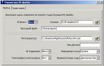
The condition of the scar on the uterus is assessed using ultrasound. Signs of insolvency are a contraindication to IVF. Stimulation protocols for fibroids Preparations for artificial insemination preparation can provoke re-growth of the tumor.
Therefore, preference is given to the following schemes:
- A short protocol is the use of a gonadotropin-releasing hormone agonist from day 2-3 of the menstrual cycle along with gonadotropic hormones.
- Long protocol - administration of a GnRH agonist from the middle of the luteal phase. Apply drugs such as Diferelin, Decapeptil, Suprefact. Enter subcutaneously in the navel.
- GnRH antagonists (orgalutran, cetrotide) are used in combination with gonadotropins.
The probability of IVF result in fibroids
According to various studies, if the node does not change the shape of the uterine cavity, is small in size and is located in the thickness of the muscles, then the pregnancy rate after artificial insemination is up to 37%. If myomectomy was performed, and after it stimulation was carried out, from 35 to 37% of women became pregnant.
The location of the node is intramural, the increase in the size of the uterus due to it reduces the frequency of pregnancies to 12% at the first attempt. During gestation, complications often develop in the form of a threatened miscarriage, bleeding, and premature birth.
Tumor recurrence within a year after treatment naturally reduces the number of pregnant women even after a long stimulation protocol.
Effect of pregnancy on fibroids
Small myoma nodes do not yet have their own system of regulation and production of hormones, so they are subject to hormonal fluctuations in the body. Myoma after IVF up to 5 cm during pregnancy decreases in size or its growth stabilizes. In some cases, after childbirth, the node is not detected.
But in about 30% of cases, pregnancy provokes an increased growth of the focus, which can increase by 2 times. Childbirth in many cases is proposed to be carried out by caesarean section.
After extraction of the fetus, a myomectomy or removal of the uterus is possible.
Yulia Shevchenko, obstetrician-gynecologist, specially for the site