Different sizes of cerebral ventricles in the fetus. Interpretation of ultrasound of the stomach of the fetus at the second screening
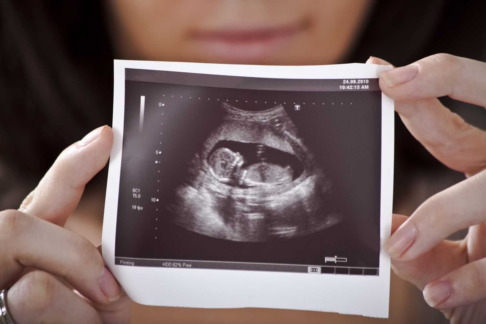
Pregnant women who have been diagnosed by ultrasound the fetus has an enlarged stomach can't find a place for themselves. They are tormented by the question of why this happened, whether it is dangerous or not and what can be done? I know this from my own work experience, when, when conducting an ultrasound examination, the size of the baby’s stomach turned out to be larger than normal. So, let’s look into this issue so that there are no misunderstandings.
One side of the coin
As a rule, enlarged stomach in the fetus is a functional state. This means that its size directly depends on what the pregnant woman ate. So, if her diet on the eve of the study was dominated by foods accompanied by increased gas formation, then the baby’s stomach becomes enlarged. What to do? It is enough to undergo a dynamic ultrasound in a week, having previously excluded from the diet the following foods the day before:
- Radish
- Peas
- Beans
- Cabbage
- Rye bread
- Milk
- Kefir
- Cottage cheese
- Apples
- Pears
- Grape
- Green.
It is best to eat during the day before the test:
- Crackers
- Porridge pudding (porridges themselves can also lead to increased gas formation).
If, upon repeated ultrasound, the size of the fetus’s stomach is normal, then there is no cause for concern.
The other side of the coin
Much less often enlarged stomach in the fetus may be a sign of an abnormal structure of his gastrointestinal tract. Most often we are talking about intestinal obstruction caused by obstruction of one or another part of the intestine. Another reason may be a mass formation in the abdominal cavity, which compresses the digestive tract. To exclude these conditions, dynamic ultrasound is performed. If, upon repeated examination, the diagnosis is confirmed, then it is necessary to give birth in a perinatal center, where it is possible to perform emergency surgery immediately after birth.
Samsung Medical Equipment
The largest microelectronics manufacturer, Samsung has maintained its flagship position in the field of personal digital technology for many years. Products under this brand have become firmly established in the life of modern people.
At the same time, the South Korean concern is a leader not only in the consumer market, but also in professional segments, in particular, in the production of innovative medical equipment.
High tech Samsung Ultrasound and X-Ray Systems open up unique opportunities for professionals. The digital image processing technologies developed by the company have been highly praised by experts.
For a long period, Professor Christoph Lees (an expert in fetal medicine) conducted research using medical equipment Samsung. “Many months of experience in our work... convinced us of the existence of fundamentally new opportunities in the field of intrauterine observation, and in particular – the skeletal system, brain and facial structure of the fetus,” writes the scientist in the authoritative academic publication “Ultrasound in Obstetrics & Gynecology” (March 2016 G.).
Samsung medical equipment is supplied to Russia by Mediays, the official dealer of Samsung Medison Co. Ltd. Over 20 years of cooperation, several thousand ultrasound scanners of various configurations have been supplied to medical institutions in our country. Since 2014, Samsung Electronics began supplying and selling digital radiographic equipment.
Through a branched dealer network "Mediays" Samsung medical equipment is supplied and serviced throughout the territory Russian Federation. The latest samples of ultrasonic and radiographic equipment from the Republic of Korea are demonstrated by the company at central and regional exhibitions. The MediaEys offices in Moscow, Novosibirsk and Yekaterinburg have showrooms where company managers give lectures to doctors and conduct practical classes on the principles of operation and design of equipment.
"Mediays" provides warranty and post-warranty service for supplied medical equipment. At the plant in the Republic of Korea, the company's engineers are trained in the installation, configuration, maintenance and repair of Samsung medical equipment. It is important to note that MediaEys service support is provided only for medical equipment officially supplied to the Russian Federation. Certified service centers operate in Barnaul, Vladivostok, Volgograd, Vologda, Voronezh, Yekaterinburg, Kazan, Kirov, Nizhny Novgorod, Perm, Pyatigorsk, Rostov-on-Don, Samara, St. Petersburg, Saratov, Tver, Ufa and Chelyabinsk.
Medical journal on echography
In Russia, the magazine "SonoAce-Ultrasound" (formerly international medical journal"SonoAce-International") appeared in 1996. The journal contains up-to-date clinical information and focused on doctors Ultrasound diagnostics, published by Mediaace and distributed free of charge to more than 3,000 medical institutions in the Russian Federation.
Subjects of the journal articles: diagnostics of the abdominal cavity, superficial organs and vessels, the use of ultrasound in obstetrics and gynecology, echocardiography, etc.
Numerous letters to the magazine confirm its relevance. The magazine's editorial board thanks readers for their attention and high ratings.
The magazine "SonoAce-Ultrasound" is sent by mail to the official addresses of medical organizations in the Russian Federation. For readers who do not have access to the printed version, the website publishes electronic version magazine in full.
All about ultrasound
Ultrasound is a medical imaging technique that began to be used more than 40 years ago. Currently, medicine can no longer imagine its existence without this diagnostic method. The areas of application of ultrasound in medicine are extremely wide. For diagnostic purposes, it is used to identify diseases of the abdominal and kidney organs, pelvic organs, thyroid gland, mammary glands, lymphatic system, heart, blood vessels, in obstetric and pediatric practice. Due to the physical properties of ultrasound, organs containing air and bone tissue are inaccessible to this method.
Every year a large number of meetings of ultrasound diagnostic specialists take place in Russia. The “Mediays” company organizes and provides sponsorship assistance in holding regional congresses of associations of ultrasound diagnostic specialists, scientific and practical conferences and seminars. The topics of the events are determined by the needs of specialists in the relevant region.
In the news section we publish reports on conferences and exhibitions of medical equipment, announcements of events with our participation.
The echogram atlas was created to demonstrate the capabilities of Samsung Medison scanners. Examples of ultrasound examinations are divided into areas of study and types of scanners, and there is a special selection of 3D echograms and videos.
All echograms of the atlas were obtained using Samsung Medison devices. The main part of the database was received from Korea, new echograms are from users of Samsung Medison scanners in Russia. The material is recommended for viewing by ultrasound diagnostic specialists.
Did you know that modern ultrasound scanners allow you to obtain three-dimensional images of objects with a resolution of up to 0.1 mm, examine small vessels and tissue textures, observe blood flow in vessels, and the movement of the walls of the heart?
What is Doppler mapping and elastography? Read the answers to these and other questions about methods of obtaining ultrasound images in the “Technologies” section.
The section contains basic concepts, how an ultrasound examination is carried out and whether it is necessary to prepare for it, what 3D ultrasound is, deciphering the examination protocol and much more.
Why is it important to assess the condition of the baby’s internal organs? The fact is that some deviations in their development are markers of chromosomal abnormalities. Such pregnant women are then recommended to undergo genetic counseling.
The timing of the screening study (16-22 weeks) was chosen so that if severe intrauterine developmental anomalies incompatible with life are detected, the pregnancy can be terminated.
Norms for the size of the fetal stomach on ultrasound and possible deviations
The fetal stomach at 16-20 weeks can have variable sizes. They are defined in millimeters. The normal sizes of the fetal stomach by week are presented in the table.
The study may reveal the following deviations:
- large or small stomach in the fetus on ultrasound;
- absence of an organ;
- presence of pseudo-content;
- slit-like stomach;
- gastric atresia in the fetus.
Causes of abnormalities in the fetal stomach
The stomach on ultrasound is defined as a round or oval anechoic formation located in the upper left parts of the abdominal cavity. Changes on its part may be signs of gastrointestinal pathology or indirect signs of other unfavorable conditions.
If The fetal stomach is not visible on ultrasound, then suggest esophageal atresia. In this case, the organ is there, but it is not filled with amniotic fluid. The absence of echoshadow may indicate oligohydramnios, and this indirectly indicates kidney pathology, chronic infections of the mother, pathology of the placenta, and gestosis. Sometimes the fetal stomach is not visualized if it is displaced from its usual place. This condition occurs with a diaphragmatic hernia.
Fluid in the stomach sometimes absent due to defects of the central nervous system, cleft lip, neuromuscular diseases.
"Pseudo-contents" of the stomach- these are various hyperechoic inclusions. They are formed when clumps of cells are ingested. Sometimes an intrauterine tumor manifests itself this way, but it is rarely diagnosed in isolation; it is accompanied by other anomalies. Swallowed blood also gives the picture of a full stomach. In this case, you need to look for the cause of the bleeding and carefully examine the placenta for abruption. Blood may appear after amniocentesis is performed.
If no pathology of the placenta or other defects is detected, then the isolated presence of “pseudo-content” is taken as a variant of the norm.
Reason enlarged stomach in the fetus There may be intestinal obstruction and obstruction. But it should be remembered that the stomach can expand to significant sizes and this is the norm. Therefore, if the size of an organ deviates from the norm, you need to look for additional symptoms:
- polyhydramnios;
- thickening of the walls of the organ;
- the vestibule is narrowed and funnel-shaped;
- no slight curvature.
Microgastria of the fetal stomach does not occur as a separate congenital disease. It is usually part of the pathology of the entire gastrointestinal tract. It can be combined with:
- violation of rotation of abdominal organs;
- absence of a gallbladder;
- abnormal position of the liver.
With microgastria, the stomach looks small, significantly smaller in size than normal, and may be cylindrical in shape.
Slit-like stomach in the fetus means its underdevelopment during onotogenesis.
Microgastria and slit-like stomach are subject to observation after detection. After the baby is born, perform plastic surgery to create a stomach from part of the small intestine. It will not be able to perform its main functions, but will keep food from directly entering the intestines. This will increase the digestion time, and the child will be able to absorb more nutrients.
Gastric atresia- a rare anomaly. This is the presence of a membrane in the pyloric or antrum. Sometimes the fusion is incomplete, there is a small hole. Like most developmental defects, there is a combination of atresia with anomalies of the larynx, esophagus, pulmonary dysplasia, ascites, and hydrothorax. Sometimes it is a sign of autosomal recessive hereditary diseases, combined with epidermolysis bullosa. Isolated pyloric atresia can be treated surgically, with a survival rate reaching 90%.
The fetus may not have a stomach. This condition is called agastria or agenesis. In the first case, the pathology is the absence of the upper part of the abdominal wall and the organs of half of the abdominal cavity. Agenesis is the absence of an organ. An extremely rare pathology, combined with multiple defects of other organs. These developmental anomalies are incompatible with life.
What to do if a fetal stomach pathology is detected by ultrasound?
Any pathologies identified during screening require repeated ultrasound diagnostics over time. It is carried out at 22 weeks. If the anomalies detected in the previous study were transient in nature, then they will disappear with repeated diagnostics. Persistent changes speak in favor of serious congenital diseases.
In case of severe concomitant malformations, the woman is offered termination of pregnancy for medical reasons. The birth of such a child is impossible; it will end in death in the first few days.
Yulia Shevchenko, obstetrician-gynecologist, especially for the site
Useful video