How to cure streptococcal infection. Test methods for streptococcal infection
The genus Streptococcus, family Streptococcaceae includes 21 species. Most often they cause diseases: S.pyogenes, agalacticae, faecalis, greening (pneumoniae).
Pyogenic streptococci
Morphology. Gr+, spherical or oval. In smears they are located in pairs or short, 6–8 cell chains. They have a capsule, but the hyaluronic acid contained in it is not antigenic. Motile, do not form spores.
Streptococci are CULTIVATED in nutrient media with the addition of glucose, serum or blood. On the surface of dense media they form small (up to 1 mm) colorless colonies, in liquid media they form bottom and wall growth, while the medium is transparent. According to the nature of growth on blood agar, they are distinguished: beta-hemolytic - a transparent zone of hemolysis forms around the colonies; alpha-hemolytic – narrow, greenish zone; non-hemolytic - the environment does not change.
They produce a number of enzymes; lactase and sucrase are of differential diagnostic importance.
AG. Based on the AG composition of the polysacchoride cell wall (substance C), streptococci are divided into 20 serogroups (designated in capital letters of the Latin alphabet A–V). Within serogroups, streptococci are divided into serovars (designated by numbers). Most causative agents of steptococcal infections belong to serogroup A.
Ig-new Fc receptors of the cell wall, lipoteichoic acids, as well as toxins and enzymes secreted into the environment during reproduction by microorganisms have antigen properties. Pathogenicity for humans is determined by the formation of toxins, extracellular enzymes and the properties of the bacterial cells themselves. The list of diseases caused by streptococci is quite large: tonsillitis, chronic tonsillitis, scarlet fever, purulent skin lesions, phlegmon, sepsis, nephritis, rheumatism, otitis media, etc. Attempts to find differential signs of streptococci, which cause diseases so diverse in clinical manifestations, were unsuccessful. Only in relation to scarlet fever streptococcus has it been established that they can secrete erythrogenic toxin; other signs are the same as other streptococci of the SEROLOGICAL GROUP A. Streptococci serovar 12, which produce cytotoxin, are considered nephritogenic.
Streptococcus serogroup IN(S. agalactia) can cause postpartum infections and sepsis in newborns, erosive stomatitis, urogenital infections in women, sepsis, and meningitis.
WITH– causative agents of respiratory infections, MPS diseases.
N And TO– isolated from endocarditis.
D(enterococci) - live in the intestines of a healthy person, cause damage to the biliary tract, and can cause endocarditis by getting a purulent-inflammatory infection into wounds. With generalization – sepsis.
Some (S.mutans, S.salivarius, etc.), which do not contain group Ag, live in the oral cavity. S.mutans is involved in the development of dental caries and periodontal disease.
Streptococci of other serogroups are rarely found in humans.
Pathogenicity factors. Adhesins are a lipid component of the lipoteichoic acid complex with cell wall proteins that ensure interaction with the epithelial membrane and colonization. Protection against phagocytosis is provided by:
1) antichemotactic factor;
2) Ig new Fc receptor(to IgG) – suppresses phagocytosis, destroys complement, causes an imbalance of immunoglobulins;
3) capsule(in serogroups A and B) – protects against phagocytes;
4) M protein, allows you to grow and multiply in human blood; cells lacking M-protein are phagocytosed without the participation of antibodies. M protein provides the same ability to penetrate microbial cells and multiply in them.
ENZYMES: hyaluronidase (spreading factor) and streptokinase (fibrinolysin) - destroys fibrin, which limits the local focus of inflammation, promoting the generalization of the process.
Streptococci SEROGROUPS A form a number of TOXINS:
O-streptolysin(heat-labile protein) - released during reproduction, causes lysis of red blood cells, destroys the membranes of other cells, as well as the membranes of lysosomes, has a cardiotoxic effect. This toxin is an antigen and stimulates the synthesis of anti-O-streptolysin.
S-streptolysin(nucleoprotein), does not have antigenic properties, causes lysis of red blood cells, destroys lysosomes, mitochondrial membranes of human cells.
Leukocidin lyses polymorphonuclear leukocytes, turns off phagocytosis.
Cytotoxins(peptides) – damage cells. One of these toxins can damage kidney tissue; it is secreted by nephritogenic strains of streptococci serovar 12.
Erythrogenic toxin (scarlet fever). Information about the formation of this toxin enters the cell with the genome of the temperate phage. The thermostable fraction of the erythrogenic toxin stimulates the HRT response.
Ecology and distribution. There are pathogens that cause diseases only in humans, humans and animals, and opportunistic pathogens for humans. They live in the oral cavity, on the mucous membranes of the upper respiratory tract, on the skin, and in the intestines. The source is patients and bacteria carriers. The route of spread is airborne. A significant part of diseases are endogenous infections that occur in people with immunodeficiency conditions.
In the environment (on household items, in dust) they can persist for several days, and withstand drying well (retain viability, but lose virulence). Sensitive to heat and disinfectants.
Immunity. The development of streptococcal infections is influenced by the state of microorganisms. Often the disease develops against the background of pre-existing sensitization (repeated tonsillitis, erysipelas, chronic infections - tonsillitis, nephritis, rheumatism). Possible involvement of autoimmune processes (rheumatism). They have hypertension that cross-reacts with the sarcolemma of the muscle fibers of the heart.
Antibodies are produced against all streptococcal biologically active substances (toxins, enzymes). Immunity after infections (except scarlet fever) is low-strength and is of a typical antimicrobial nature (to the M-antigen). Antibodies to enzymes and streptococcal toxins have practically no protective properties. The level of sensitization intensity is checked in allergy tests.
Laboratory diagnostics. The material for BACTERIOLOGICAL research is mucus from the pharynx, pus, wound discharge, blood, etc. Isolated pure cultures are identified and their basic properties are determined: morphology, hemolytic activity, sensitivity to antimicrobial drugs. SEROLOGICAL DIAGNOSTICS - detection of antibodies to toxins and enzymes. In rheumatism, there is an increase in the titers of anti-O-streptolysins, anti-DNase, and antihyaluronidase in paired sera.
Prevention and treatment. Specific prevention has not been developed. To prevent chronic streptococcal infections associated with the persistence of the pathogen and the formation of L-forms, antibiotic therapy is used. For children who have had repeated tonsillitis, scarlet fever, dispensary observation is established (prevention of rheumatism). Serogroup A streptococci are highly sensitive to penicillin (bactericidal action); resistance to penicillin is not acquired. Sulfonamides have a bacteriostatic effect on streptococci. Microorganisms easily acquire resistance to them.
Occupies a special place SCARLET FEVER– acute infectious disease, pathogen – hemolytic streptococcus group A of any serovar, capable of producing erythrogenic toxin.
Pathogenesis. Scarlet fever is a highly contagious disease that occurs cyclically with changing symptoms. At stage 1, the effect of the erythrogenic toxin is manifested (intoxication, sore throat, pinpoint rash on a hyperemic background).
2nd period is accompanied by complications as a result of the action themselves streptococci (purulent lymphadenitis, mastoiditis, otitis), because antimicrobial immunity is not expressed, and infection with streptococci of other serovars, for which there are no corresponding antibodies.
Immunity. Unlike other streptococcal infections, strong antitoxic immunity remains, because erythrogenic streptococcal toxin all serovars AG is identical. Antimicrobial immunity type-specific and does not protect against the occurrence of other streptococcal diseases.
The intensity of antitoxic immunity to erythrogenic toxin is checked by intradermal tests. In the absence of immunity, the smallest dose of the toxin causes redness and swelling of the skin. If there are antitoxins in the blood, there is no reaction to the introduction of the toxin.
Streptococcus pneumoniae (PNEUMOCOCCUS). S.pneumoniae - causes pneumonia - pneumonia, which is explained by the specificity of adhesins that interact with lung cell receptors.
Morphology, physiology. They have an elongated shape, reminiscent of a candle flame. They are arranged in pairs, each pair is surrounded by a capsule. Under the capsule there is an M protein, which is similar in properties to S. pyogenes, but has its own antigen specificity.
On solid nutrient media to which serum or blood is added, pneumococci grow, forming small colonies surrounded by a greening zone (on blood agar). In liquid media they produce uniform turbidity.
BH activity is moderate - they break down a number of carbohydrates and form hyaluronidase, muromidase, peptinase.
Have 3 main AG: cell wall polysaccharide antigen, capsular antigen and M protein. Based on capsular hypertension, they are divided into 84 serovars.
Ecology and distribution. They live in the upper respiratory tract of humans, enter the lower respiratory tract, and with congestion in the lungs, decreased activity of SIgA and macrophages, and destruction of surfactant, endogenous pneumonia occurs. Infection by airborne droplets.
Outside the body, pneumococci quickly die. They cannot withstand heating or disinfection. Dried sputum can persist for a long time. Sensitive to penicillin, macrolides.
Pathogenicity. They produce hemolysins and leukocidin, which damage tissue cells. M protein and capsule provide adhesion ability and resistance to phagocytosis. Released enzymes play a large role in the development of the pathological process: peptidase breaks down SIgA hyaluronidase promotes the spread of microorganisms in tissues. When exposed to toxins and enzymes, macrophages under surfactant can leave the “defense line”. Generalization of the process is possible, which more often occurs in young children and the elderly. In these cases, meningitis and sepsis occur.
Immunity. Type-specific and unstable, this explains the re-occurrence and the possibility of transition to a chronic form.
Laboratory diagnostics. To isolate pathogen cells, it is necessary: an optimal nutrient medium for reproduction, cultivation conditions, and correct collection of the test material. The isolated cells are identified by a number of characteristics and differentiated from viridans (alpha-hemolytic) streptococcus and enterococci. Pneumococci with a capsule are subjected to serological typing and the sensitivity of microorganisms to antimicrobial drugs is determined.
Prevention and treatment. Specific prevention has not been developed. In each particular case, nonspecific measures are important aimed at preventing the possibility of an endogenous infection: in patients who are forced to lie down for a long time, who are on hormonal therapy, radiation therapy, and when the general resistance of the body decreases, natural defense mechanisms are stimulated (diet, vitamin supplementation, strengthening ventilation of the lungs by massage and other influences). Penicillin and macrolide antibiotics are used to treat pneumonia.
ENTEROCOCCI. S.faecalis (fecal streptococci, enterococci) are inhabitants of the intestines of humans and warm-blooded animals. Are part of the group D .
Morphologically- These are spherical or oval bacteria; when dividing, they form pairs or short chains. Polymorphic, some strains are motile, have 1–4 flagella.
Fermentation of individual carbohydrates is a variable feature.
Ecology and distribution. Enterococci are more resistant to factors environment than other streptococci. They can withstand heating up to 60°C for 30 minutes and are able to multiply in media with 6.5% NaCl, 40% bile, at pH 9.5–10.0. Potassium tellurite, sodium azide, bile salts, crystal violet, nalidixic acid, as well as penicillin and neomycin do not inhibit the growth of enterococci, which is used to create selective culture media.
Pathogenesis. They are capable of multiplying in food products; when contaminated food is consumed, foodborne illness develops. It is most often caused by proteolytic variants.
Purulent-inflammatory processes usually occur sluggishly, chronically. More often, it is not a monoinfection that occurs, but a mixed one, in association with Escherichia coli, Proteus, and staphylococci. Hemolytic variants of S.faecalis are isolated from pus, discharge from wounds, and the upper respiratory tract, where pathological processes are localized. Most strains of enterococci isolated from patients turn out to be resistant to penicillin, neomycin, and have pathogenicity enzymes - coagulase, hyaluronidase, DNAase, fibrinolysin , proteinase, muromidase. When subcultured in the laboratory, these enzymes usually cease to be released
> Examination for streptococcal infection
This information cannot be used for self-medication!
Consultation with a specialist is required!
There are several types of streptococci that cause inflammatory processes in the human body. Group B beta-hemolytic streptococcus is especially dangerous. This bacterium lives in the nasopharynx, gastrointestinal tract, and vagina. There are three serovars (subspecies). Streptococci serovar 1a and III live mainly in the respiratory tract and central nervous system. They can cause meningitis and pneumonia in newborns, so pregnant women at 35 weeks are prescribed tests to identify these microorganisms. Group A beta-hemolytic streptococcus lives primarily on the skin and mucous membranes. Causing inflammation and producing many toxins (hemolysin, streptolysin, etc.), it can cause tonsillitis, pharyngitis, as well as invasive diseases (myositis, endocarditis, etc.), scarlet fever and even toxic shock.
Among the laboratory methods for identifying streptococci, PCR testing (detection of pathogen DNA) is used. The material for the study is blood plasma, scrapings of epithelial cells of the oropharynx, saliva, sputum, washings and fluid removed from the lungs (lavage). The choice of the type of biomaterial for research is made by the doctor depending on the expected colonization of streptococcus.
To confirm the streptococcal nature of the disease, effectively prescribe treatment and confirm recovery, culture of material is carried out to detect beta-hemolytic streptococci, followed by determination of their sensitivity to antimicrobial drugs.
Also, special antigen tests are performed to identify group A and B streptococci. To detect streptococcus A using this method, a swab from the oropharynx is taken from the patient for analysis.
Indications for testing for streptococcus
Doctors prescribe a blood test to detect streptococcal DNA for septic conditions, meningitis, and acute stages of pneumonia. PCR testing of scrapings of the epithelium of the oropharynx, sputum, washings and lavage fluid is carried out for prolonged wet cough accompanied by fever, bronchitis, tonsillitis, pharyngitis, rhinosinusitis. Tests for streptococcus are indicated for patients with bronchopulmonary diseases due to reduced immunity, pregnant women, medical workers for registration of a personal medical record.
Obstetricians-gynecologists, pediatricians, therapists, neurologists, pulmonologists, and surgeons prescribe tests to detect the presence of streptococcus. Research is carried out both in multidisciplinary hospitals and in many paid laboratories.
Preparing for the study
It is advisable to get tested before starting antibiotic treatment. Collection of saliva, sputum, washings, lavage fluid and material from the oropharynx should be carried out before diagnostic and therapeutic measures are carried out in these areas. You should drink and eat no later than 2–3 hours before collecting material from the oropharynx.
Interpretation of test results
The laboratory issues the results of the PCR test and antigen tests for the presence of group A and B streptococci in the form of “detected” or “not detected”. A negative result most often indicates the absence of infection, less often - an insufficient concentration of the pathogen or its antigens in the biomaterial sample. Normally, neither streptococcal DNA nor its antigens should be detected in the tissues examined. However, healthy people can also be carriers of streptococci.
If the growth of beta-hemolytic streptococci of groups A and B is detected during culture, the doctor indicates the type of microorganisms, the number of colonies grown and their sensitivity to certain drugs. Normally, there should be no growth of streptococci.
For local drip streptococcal infections, the material for research is sputum, nasopharyngeal mucus, pus, rinses, wound discharge; for common forms of the infectious process - blood and urine.
For laboratory analysis, microscopic, bacteriological and serological diagnostic methods are used.
The purpose, features and diagnostic value of microscopic examination are the same as for staphylococcal infections.
1. BACTERIOLOGICAL STUDY
To isolate a pure culture of streptococci, it is important to create an optimal nutrient medium, since streptococci have special requirements for it. They need a significant amount of carbohydrates and native protein. Therefore, along with the generally accepted sugar MPB, blood MPA, milk-salt MPA and MPB (see recipes above), ascitic and serum media are used for streptococcal infections.
ASCITICA MPB and MPA are prepared with the addition of reporting fluid obtained sterilely from the abdominal cavity of medical and surgical patients. The liquid is heated for 3 days at +56-58 °C for 1 hour, sterilized by filtration through a Seitz filter or 40% glycerol is added and stored in the cold. To prepare ascites broth and ascites agar, 1 part of the liquid is mixed with 2-3 parts of MPB (or Hottinger broth) or melted and cooled MPA.
WHEY MPB is prepared from simple fresh meat-peptone broth with a pH of 7.6, to 1 part of which 2 parts of fresh human or horse serum are added. The serum is inactivated at + 56 °C for 30 minutes before being added to the medium.
When drip streptococcal infections are complicated by sepsis, blood culture is also necessary. For bacteriological examination of blood, E. G. Kassirskaya recommends the complex use of three types of nutrient substrates, inoculated at the rate of 1 part of pathological material per 10-15 parts of the medium. The latter uses 0.2% semi-solid agar with 10% ascitic fluid, Levinthal broth with blood and Kitt-Tarozzi liver medium.
FOR LEVINTHAL BROTH, prepare the following components separately: No. 1 - to 100 ml minced meat add 300 ml of distilled water and 10 ml of normal soda solution; No. 2 - 0.5 g of pancreatin is dissolved in 20-30 ml of water with 2 ml of 1 N soda solution and 10 ml of chloroform; No. 3 - buffer solution of sodium phosphate in distilled water (diluted 8:1000). Using a HCl solution, the pH is adjusted to 5.6-6.
On the first day, mixture No. 1 is incubated in a thermostat at + 37 °C for 1-2 hours, solution No. 2 is added to it, mixed and kept under the same conditions for another 24 hours. The vessel with the medium is shaken periodically. After this, take equal amounts of meat pulp and buffer solution No. 3. Boil and filter. Set pH to 7.2-7.4. They boil again. Pour into test tubes and sterilize for 2 days in a row for 30 minutes with running steam.
KITT-TAROZZI MEDIUM is made from beef liver or meat. The latter are cut into pieces, weighed, poured with a triple amount of MPB (pH-7.4-7.6) and boiled for 30 minutes. Then the broth is filtered, and the liver pieces are washed with tap water. Next, test tubes with 3-4 pieces of liver, filled with 7-8 ml of filtrate and a layer of vaseline oil, are sterilized under a pressure of 1 atm. within 30 minutes.
The incidence of streptococci will increase when using SEMI-LIQUID GAROZZI AGAR: 0.3-0.5% glucose and 0.1-0.15% agar-agar are added to Martin broth (pH-7.6-7.8). Place pieces of liver or boiled meat into sterile test tubes, add 9 ml of medium and sterilize at +120 °C for 30 minutes.
Viridans streptococcus, isolated in septic endorcarditis, develops very slowly. In this regard, blood cultures are kept in a thermostat for 2-3 days.
In some cases, it is not possible to isolate a streptococcal culture with extensive aeration. The use of anaerobiosis is more successful. To create the latter, you can use three simplest methods.
I. THE MATERIAL TO BE TESTED IS SEEDED INTO A TEST TUBE with 0.25% glucose broth and quickly sucked into sterile Pasteur pipettes, the ends of which are immediately sealed over a burner flame. The pipettes are installed vertically in the thermostat. After 24 hours, the lower ends of the pipettes are broken off (streptococci grow only at the bottom), the first drops are used for microscopy and further isolation of a pure culture of the pathogen.
2. CULTIVATION OF CROPS IN AN ATMOSPHERE SATURATED WITH CARBON DIOXIDE. The required concentration of CO 2 is obtained by adding first 1 g of bicarbonate of soda per 1 liter of volume to a desiccator loaded with test tubes, and then, from the same calculation, 8-9 ml of 10% H 2 SO 4 or HCl.
3. PRETTY SIMPLE and less effective is the following technique: place a lit candle at the bottom of a loosely closed desiccator. It burns for 1-3 minutes and goes out. At the end of the first or second procedure, the desiccators are covered with lids, the edges of which are greased with Vaseline and placed in a thermostat.
Isolation of pure culture
The biochemical activity of streptococci is variable and its determination has no diagnostic value. The study of streptococci in this regard is used only for differentiation from enterococci (Table 1).
| Table 1. Differentiation of streptococci from enterococci | ||
| Sherman criteria for distinguishing group A streptococci (true) from group D (entrococci) | ||
| Tests | Groups | |
| Group A (streptococci) | Group D (enterococci) | |
| Chain length | long (5-12 links) | short (1-2 links) |
| Growth in salt MPA from 6.5% | + | - |
| Growth on the bile-blood MPA of D. E. Belenky to P. N. Popova | - | + |
| Growth on milk with methylene blue | - | + (reduction) |
| Growth on MPB with pH - 9.6 (in the presence of 0.05 M Na 2 CO 3 solution) | - | + |
| Sensitivity to penicillin | + | - |
| Heat resistance at +60 °C for 30 minutes. | - | + |
The composition of differential diagnostic media used for this purpose is as follows.
- BILE AND BILE-BLOOD MPA D. Z. Belenky and N. N. Popova are prepared from melted and filtered 3% MPA with any broth base. To 60 ml of this MPA, add 40 ml of native filtered bile, pour it into bottles and sterilize at a pressure of 1 atm. 30 minutes. To prepare blood agar, 5% defibrinated blood is added to this bile MPA.
- MILK WITH METHYLENE BLUE is prepared from skim sterile milk, to 100 ml of which 2 ml of a 10% aqueous solution of methylene blue is added.
DETERMINATION OF STREPTOCOCCUS VIRULENCE
To prove the pathogenicity of streptococci, hyaluronidase activity, detection of streptokinase or fibrinokinase, plasmacoagulase, leukotoxic effect of streptococcus, and the presence of hemolysin are important. The determination of these indicators is carried out using the methods described above, but the detection of hemolyzing activity of streptococcus is better done on media with human blood.
DETERMINATION OF LEUKOCIDIN. Citrate blood of a person or any animal is taken, centrifuged, the upper yellow layer of leukocytes is sucked off with a pipette, transferred to another tube and a 2-5% leukocyte suspension is prepared. The latter is poured into 1-1.5 ml into narrow test tubes. 1 loop of 1-2 billion daily culture of streptococcus is also added here and placed in a thermostat at +37 °C for 1 hour. After incubation, smears are made from the leukocyte-microbial mass (similar to smears from whole blood), dried, and fixed for 15 minutes. in Nikiforov’s mixture, stained for 45-60 minutes according to Romanovsky-Giemsa, microscopically. Massive destruction of leukocytes indicates the presence of leukocidin.
DETERMINING THE SENSITIVITY of isolated cultures to medicinal substances is carried out using generally accepted methods.
SEROLOGICAL TYPING of detected streptococci after their isolation is required only for special epidemiological purposes and is rarely used.
II. SEROLOGICAL METHOD FOR DIAGNOSTICS OF STEPTOCOCCAL INFECTIONS
Streptococcal virulence enzymes (hyaluronidase, fibrinokinase, plasmacoagulase) and their toxins (for example, hemotoxin) are powerful antigens, in response to which the corresponding antibodies are produced: antihyaluronidase, antistreptokinase, antistreptolysin, etc. By detecting these antibodies, the disease and phase can be diagnosed development of the infectious process.
DETERMINATION OF ANTISTREPTOLYSIN (ANTIHEMOLYSIN)
Streptolysin is a type of hemotoxin. Its presence is checked on red blood cells. In response to the action of this antigen, antibodies are formed in the body that can neutralize its hemolyzing activity. When antistreptolysin is detected, the following is needed: patient serum with antistreptolysin (antibodies); streptolysin (purified), standard, lyophilized; 5% suspension of rabbit, sheep or human red blood cells; phosphate buffer for diluting serums and preparing a suspension of erythrocytes: dissolve 7.6 NaCl, 3.17 g of KH 2 P0 4 and 1.81 g of Na 2 HPO 4 in 1 liter of distilled water, add concentrated NaOH dropwise and adjust the pH to 6.5- 6.7. The buffer solution is stored in a refrigerator at -4 °C for 2-3 weeks.
The determination of antistreptolysin consists of two stages: the first is the establishment of the titer and working dose of standard streptolysin, the second is the identification and quantitative determination of antistreptolysin. Schemes for their implementation are given below.
| Scheme for determining the working dose of standard streptolysin | |||||||
| Components in ml | Test tubes | ||||||
| 1 | 2 | 3 | 4 | 5 | 6 | 7 | |
| (red blood cell control) | |||||||
| Streptolysin | 0,6 | 0,7 | 0,8 | 0,9 | 1,0 | 1,1 | - |
| Buffer solution | 0,9 | 0,8 | 0,7 | 0,6 | 0,5 | 0,4 | 1,5 |
| Red blood cell suspension | 0,5 | 0,5 | 0,5 | 0,5 | 0,5 | 0,5 | 0,5 |
| Place in thermostat for 15 minutes at +37 °C, shake, then return to thermostat for 30 minutes. | |||||||
| Result | - | - | - | - | hemolysis | hemolysis | - |
The titer and working dose of streptolysin is considered to be its minimum amount that gives clear hemolysis of red blood cells. In this example they are 1.0 ml.
IN lately A standard lyophilized stretolysin is produced, the bottle with which and the attached instructions indicate the method of diluting the drug to obtain a working dose. This streptolysin provides good repeatability of results.
| Scheme for setting up a reaction for determining antistreptolysin | ||||||
| Components in ml | Test tubes | |||||
| 1 | 2 | 3 | 4 | Control | ||
| 5 | 6 | |||||
| streptolysin | red blood cells | |||||
| Buffer solution | 0,5 | 0,5 | 0,5 | 0,5 | 0,5 | 0,5 |
| Patient serum | 1:50 | 1:100 | 1:200 | 1:400 | 1:800 | - |
| Streptolysin at work. dose. | 1,0 | 1,0 | 1,0 | 1,0 | 1,0 | - |
| Shake, thermostat at + 37 °C for 15 minutes. | ||||||
| Red blood cell suspension | 0,5 | 0,5 | 0,5 | 0,5 | 0,5 | 0,5 |
| In the thermostat for 45 minutes at +37 °C with periodic shaking. | ||||||
| Result | - | - | hemolysis | hemolysis | hemolysis | - |
Antistreptolysin was detected in this serum sample of the examined patient at a titer of 1:200.
DETERMINATION OF ANTI-HYALURONIDASE
The detection principle is based on recording the destructive effect of the enzyme hyaluronidase on the hyaluronic substrate. Antibodies in the patient's serum directed against this enzyme neutralize it and hyaluronic acid remains unchanged.
Necessary reagents: patient serum with antibodies (antihyaluronidase); hyaluronic acid extract with a known working dose (method described above); hyaluronidase (chemically pure drug); 15% acetic acid - indicator; saline solution.
The determination of antihyaluronidase consists of three stages: the first - determining the titer and working dose of hyaluronic acid, the second - hyaluronidase, the third - identifying the presence and titer of antihyaluronidase.
Titration of hyaluronic substrate is described above. The titer and working dose of standard hyaluronidase corresponds to the minimum amount that can destroy hyaluronic acid taken in a working dose.
After determining the hyaluronidase titer, the antihyaluronidase titer is determined.
| Scheme for setting up the reaction for determining the titer of serum antihyaluronidase | ||||||||
| Ingredients in ml | Test tubes | |||||||
| 1 | 2 | 3 | 4 | 5 | 6 | Control | ||
| 7 | 8 | |||||||
| hyaluronidase | hyaluronic acid | |||||||
| Phys. solution | 0,5 | 0,5 | 0,5 | 0,5 | 0,5 | 0,5 | 0,5 | 0,7 |
| Patient serum diluted 1:25 | 1:50 | 1:100 | 1:200 | 1:400 | 1:800 | 1:1600 | ||
| Hyaluronidase in a working dose | 0,2 | 0,2 | 0,2 | 0,2 | 0,2 | 0,2 | 0,2 | - |
| In the thermostat at + 37°C for 30 minutes. | ||||||||
| Hyaluronic acid in working dose | 0,3 | 0,3 | 0,3 | 0,3 | 0,3 | 0,3 | 0,3 | 0,3 |
| In the thermostat at +37 °C for 30 minutes. 15% acetic acid, 2-3 drops per test tube. | ||||||||
| Results | clot | clot | clot | - | - | - | - | clot |
In this example, the titer of antihyaluronidase in the patient’s blood serum is 1:200. This amount of antibodies at the specified serum dilution still has a neutralizing effect on hyaluronidase and prevents the destruction of hyaluronic acid. Its integrity is registered by the formation of a clot after adding an indicator - a 15% acetic acid solution.
- Dyachenko S.S. Microbiological methods for diagnosing infectious diseases. State Medical Publishing House of the Ukrainian SSR, p. 313.
- Guide to microbiology, clinical practice and epidemiology of infectious diseases. Ed. "Medicine", vol. VI, section. VI, pp. 440-474, 489-500 (enterococci).
- Guide to microbiological diagnosis of infectious diseases, ed. K. I. Matveeva and M. I. Sokolova, p. 450.
- Sachkov V.I. Immunological methods for studying rheumatism and other collagenous diseases. Medgiz, M., 1962.
- Ioffe M. F. Immunology of rheumatism. Medgiz, M., 1961.
Source: Motavkina N.S., Pyanova R.E. Microbiological diagnosis of some droplet infections and toxoplasmosis. Methodological development for students. VSMU, 1973
Infectious diseases are caused by a variety of microorganisms, among which bacteria occupy a significant proportion. In particular, streptococci. These are spherical, gram-positive, anaerobic bacteria that live mainly on human mucous membranes. An analysis for streptococcal infection is prescribed in many cases; this is the only means of identifying the pathogen if a bacterial cause of the disease is suspected.
In what cases is diagnosis carried out? First of all, for sore throat and pharyngitis, some other diseases of the upper respiratory tract. Bacteria are detected by infectious diseases intestines, in some cases, with infection of soft tissues, bones, sepsis.
In addition, laboratory diagnostics of streptococcal infections is prescribed if bacterial carriage is suspected; scrapings are taken from employees of medical institutions.
In case of nosocomial infection, testing for streptococcus is mandatory, along with identifying other pathogenic bacteria.
Tests are also done during antibiotic treatment to assess the effectiveness of therapy. An examination may be prescribed before hospitalization, as a preventative measure.
Diagnostic methods
Today, various studies can be used to identify the pathogen. The most commonly used methods are:
- bacteriological;
- serological;
- rapid testing;
- PCR diagnostics.
Each of these methods has its own specifics and capabilities, although in terms of accuracy they are all equally informative.
Bacteriological method
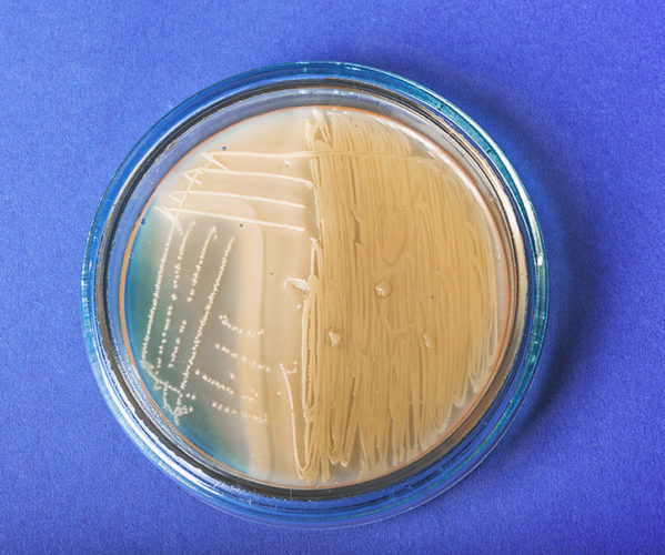
It is also called sowing. During the study, biomaterial taken from the patient is placed in a Petri dish with a nutrient medium - a mixture of carbohydrates and native protein. Three are used for sowing different types substrate, each of which is suitable for a specific type of bacteria.
Next, the Petri dishes are placed in a special thermostat and kept there for a certain time. After this, the result is studied - colonies of bacteria are formed on the nutrient media. As a rule, streptococci form round colonies that can be matte, shiny or slimy - depending on the group of bacteria.
Such an analysis is carried out to isolate an individual pathogen from a group - after all, each biomaterial contains many microorganisms, pathogenic or not. It is clear that if there are the most colonies of streptococcus, then the disease is caused by it.
A type of bacteriological analysis is anaerobiosis. The biomaterial is placed in a test tube with nutrient broth, but without oxygen. Under these conditions, only anaerobic bacteria, which include streptococci, can develop. The advantage is that all aerobic microorganisms are immediately cut off, and this increases the accuracy of diagnosis and simplifies it several times.
Serological method
It is also used to determine infection with streptococcus, but what is taken into account is not the amount of the pathogen, but the amount of antibodies to it. As you know, the human body produces antibodies to any microorganism that eliminate the danger. The more there are in the blood, the more bacteria there are. This is what serological testing is based on.
In the case of streptococcus, the amount of antibodies to streptolysin type O, which is secreted by these bacteria, is determined. It is this substance that causes the destruction of human body cells.
Depending on the detected number of antibodies, you can understand not only the cause of the disease, but also the severity of the inflammatory process - the more there are, the more streptococci in the body.
PCR
The abbreviation stands for polymerase chain reaction. This is a relatively new method that is renowned for its accuracy. The study is based on identifying specific streptococcal DNA fragments. If the study reveals a gene sequence that is characteristic of this particular pathogen, there is no doubt.
How is this possible? PCR makes copies of DNA many times using the enzyme DNA polymerase. In this case, it is possible to identify the pathogen at the earliest stages of the disease and determine its quantity in the biomaterial. The method is truly informative, in this regard it is superior to all others.
The disadvantage of polymerase chain reaction is the higher cost of the analysis, but this is offset by exceptional accuracy, which is critical in many cases.
Quick diagnosis
If a test for streptococcus needs to be done quickly, and this is required in many cases, the best option would be to take a smear followed by rapid diagnosis. It allows you to make a diagnosis within 5 minutes and does not cause any serious inconvenience.
How and with what help is rapid testing carried out? Biomaterial is taken from the patient (done in the throat with a cotton swab). After this, the stick is dipped into a test tube with a special solution and left in it for 1 minute. Next, the stick is pulled out, put into a test tube, after which the test strip is lowered there for 5 minutes. After this time, the result is evaluated.

This test is not used everywhere, although it is convenient and allows you to diagnose an infection right in the doctor's office. The reason is that test strips can give false results. Therefore, even if diagnostics have been carried out, bacterial culture is still done to clarify the diagnosis.
Biomaterials used
Microbiological diagnosis of streptococcal infections can be carried out using:
- sputum;
- nasopharyngeal mucus;
- pus;
- washings from mucous membranes;
- discharge from the wound;
- vaginal smear;
- sperm;
- urine;
- feces
The specific type of biomaterial is determined by the presumptive diagnosis - if there is a suspicion of inflammation of the bladder, then a urine test is performed, and if the baby has obvious signs of an intestinal infection (loose stools, high temperature), then the bacteria are determined in the feces.
Interpretation of analyzes
No matter how old the patient is, be it an adult, a teenager or infant, there is a certain norm for the content of streptococcus in each type of biomaterial.
The most common diagnosis of streptococcal infection is bacterial culture. Its results are assessed using the counting method. Normally, the bacterial content should not exceed 10 to the 4th or 10 to the 7th degree, no matter whether the concentration of streptococci in saliva, a swab from the throat or the cervical canal is calculated.
Pathology is indicated by a concentration higher than 10 in 7. It does not matter whether streptococcus Agalactica or any other is sown, for example, streptococcus Viridans. The more the number of bacteria differs from grade 7, the more pronounced the pathological process.
You should focus on the following reference values:
- negative result (no bacterial growth) – no infection;
- positive result with a value of up to 10 in 7 – asymptomatic carriage;
- a positive result with a value of more than 10 in 7 – acute infection.
It must be taken into account that the results of the study are always influenced by antibacterial therapy, therefore, if the patient took any drugs, it is worth knowing about it. However, this is why bacterial culture is used to determine the effectiveness of antibiotics and the sensitivity of bacteria to them.
Is treatment necessary?
Of course, if there is inflammatory process, therapy is necessary. In the case of carriage, which occurs without any symptoms, the need to take antibiotics is determined by the doctor based on the number of microorganisms identified - if it does not exceed the above indicators or is much lower, you can do without treatment.
In cases where the number of bacteria approaches the upper limit of normal, therapy is carried out, especially if there is a suspicion of decreased immunity or there are risks of developing an infection.